
Enzyme-linked immunosorbent assay (ELISA) is extremely sensitive test
that is used to detect specific antibodies or antigens. The direct
ELISA is a test for the detection of antigen.
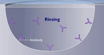
In this procedure, a known antibody is adsorbed to the inside of the
well in a microtiter plate. After rinsing to remove excess antibodies,
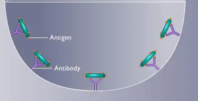
the sample suspected of containing antigen is added.
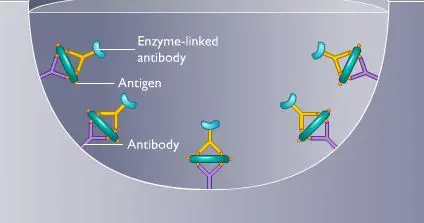
Next, an enzyme-linked antibody that can react with the antigen is added. If antigen is present in the well, the enzyme-linked antibody binds to it and is retained.
The colorless substrate for the enzyme is then added. Development for color indicates the presence of the antigen.
Next, an enzyme-linked antibody that can react with the antigen is added. If antigen is present in the well, the enzyme-linked antibody binds to it and is retained.
The colorless substrate for the enzyme is then added. Development for color indicates the presence of the antigen.
- Antigen specific antibody (Ab1) is attached to a solid phase surface.
(i.e. Antibody directed against specific infectious agent in question is firmly fixed to a solid matrix, either the inside of the walls of microdilution tray or the outside of a spherical plastic or metal bead or some other solid matrix) - Test Specimen is added
(if antigen is present in the fluid to be tested, stable antigen-antibody complexes is formed; unbound antigen is thoroughly removed by washing) - Second antibody against the antigen being sought is then added to
the system. This antibody has been complexed to an enzyme such as
alkaline phosphatase or horseradish peroxidase.
- If the antigen is present on the solid matrix, it binds the second antibody, forming a sandwich with antigen in the middle. Unbound labeled antibody is removed by washing.
- A chromogenic enzyme substrate is added. The hydrolysis of the enzyme substrate causes the color change and completes the action.
- The color developed is proportional to the amount of antigen present in the test specimen.


0 Comments:
Post a Comment