Showing posts with label Genetics. Show all posts
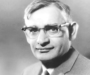
Har Gobind KhoranaBorn - 9 January 1922
Achievements - Har Gobind Khorana is an American molecular biologist, who was born to an Indian Punjabi couple. For his work on the interpretation of the genetic code and its function in protein synthesis, he was awarded the Nobel Prize in the year 1968. 

Har Gobind Khorana is an American molecular biologist born on 9 January 1922 to an Indian Punjabi couple. For his work on the interpretation of the genetic code and its function in protein synthesis, he was awarded the Nobel Prize in the year 1968. This award was, however, also shared by Robert W. Holley and Marshall Warren Nirenberg. The very same year, he received another award 'Louisa Gross Horwitz Prize' along with Nirenberg that was presented to them by the Columbia University.
 

Read this biography to learn more about Har Gobind Khorana, who became a citizen of the United States of America in the year 1966. In present times, he's residing at Cambridge in Massachusetts, United States as a part of the MIT Chemistry faculty. Har Gobind Khorana was also the first to produce oligonucleotides, which is chains of nucleotides. He was also the first person to segregate DNA ligase, an enzyme that connects sections of DNA together. 

These custom-designed portions of artificial genes are extensively used in biology labs for sequencing, cloning and engineering new plants and animals. This invention by Dr. Khorana has become automated and commercialized so that anyone now can order a synthetic gene from any of a number of companies. Thus, this is the history of the life of Dr Har Gobind Khorana as a biologist.

The following points highlight the top five cases on colour blindness in humans.
Case # 1. Of what type will be the children with reference to colour blindness, when a man is colour-blind and his wife is normal?
Solution:
The cause of the colour blindness is the presence of recessive (c) gene on the X chromosome.
Because man is colour-blind (Xc Y) and his wife is normal (XX), following will be the results while cross­ing:
Following will be the results after fertilization:
(i) XXC, i.e., normal but carrier daughter.
(ii) XY, i.e., normal son.
Results:
No child will be colour-blind.
Case # 2. When a haemophilic male is mated with a heterozygous haemopbilic female, what haemophilic proportion will be resulted in each sex?
Solution:
Haemophilia is a disease that causes delayed clotting of blood. It is due to a recessive gene ‘h’, located on X chromosome.
Haemophilic gene is represented by ‘h’
Haemophilic male = XhY
Heterozygous haemophilic female = XhX
Results:
One haemophilic daughter
One carrier daughter
One haemophilic son
One normal son.
Case # 3. When a haemophilic male is mated with a ho­mozygous non-haemophilic female-What will be the result?
Solution:
Haemophilic male = XhY
Homozygous non-haemophilic female = XX
Result:
A ratio of 2 (carrier daughter) and 2 (Normal son) will be produced.
Case # 4. Of what type will he the children with reference to colour blindness, when a woman is colour-blind and her husband is normal?
Solution:
Colour-blind woman = XCX
Normal man = XY
Result:
In such a case one normal and one colour-blind son, and one normal and one carrier daugh­ter would be resulted.
Case # 5. When both the parents are colour-blind, can they produce a normal daughter?
Solution:
Results:
The above results indicate that the colour-blind gene (c) is passed to both the X chromosomes of the daughter and so no normal daughter can be produced.

1. Structure of Plasmid:

Plasmids are small, circular, double stranded, non-chromosomal DNA molecules. In most plasmids the two ends of the double stranded DNA molecule, that make up plasmids covalently bond together forming a physical circle.
Some plasmids, however, have linear DNA. Their size vary from 1 to over 1000 kbp and contain between 5-100 non-essential genes. They play no role in the in-viability and growth of bacteria and hence called dispensable auto-nous elements.

2. Types of Plasmid:

On the basis of function, there are five types of plasmids:
1. Fertility F-plasmids:
Contain tra genes, capable of conjugation.
2. Resistance Plasmids (R-factor):
Contain genes that can build a resistance against antibiotics or poisons and help bacteria to produce pili. (Historically known as R-factors before the nature of plasmid was understood).
3. Col. Plasmid:
Contain genes that code for bacteriosins, proteins that can kill other bacteria.
4. Degenerative Plasmids:
Unable digestion of substances e.g., toluene, salicylic acid.
5. Virulence Plasmids:
Turns the bacteria into pathogens.

3. Transfer of Plasmids:

Plasmids can be transformed from one bacterium to another by transformation and gene transduction. The most common mechanism of plasmid transfer is conjugation. The plasmids which are transferred from cell to cell are known as conjugative plasmids.
They contains genes coding for proteins involved in both DNA transfer and forming of mating pairs. Plasmids can be considered part of the mobilome because they are often associated with conjugation, a mechanism of horizontal gene transfer.

4. Reproduction in Plasmid:

Plasmids replicate independently of the host chromosome, but some plasmids called episome (def) are able to insert or integrate the host cell chromosome, then their replication is integrated by the chromosome.

Co -Transformation:

It is obvious that if the exogenous DNA that enters a recipient bacterial cell, contains known marker genes, say x, y and z, then these genes appear in the trans-formants, provided the segment or segments containing these genes are successfully integrated into the host chromosome. When x and y or x and z or y and z appear in the same trans-formant, the phenomenon is called co-transformation and the particular trans-formant is called a co-trans-formant.
Naturally, the probability of co-transformation and hence the frequency of co-trans-formants depend on the relative distance between the pair of marker genes. For detecting co-transformation, the recipient bacterium must have the corresponding recessive genes, x ,y and z, because only then the presence of the x, y and z genes can be detected. For convenience, the x, y and z genes may be represented as x+, y+and z+.
Co-transformation frequency may be used for preparing gene-maps. Thus, if it is observed that xy+ trans-formants appear more frequently than x+z+, it can be concluded that x+ and y+ are closer to each oilier than x+ and z+.
As transforming DNA generally consists of fragments, a particular fragment may or may not contain a marker gene. If the fragment taken up by a cell does not contain any marker gene, there will be no transformation of the marker genes, although other genes not taken into consideration may be present. If the fragment taken up by the recipient contains a marker x+ or y+, or both markers x+ and y+, the trans-formants may have the genotypes, x+x-, y+y- or x+x-y+y. Only the last genotype represents a co-trans-formant.
Now, if the probability of x+x and y+y trans-formants in the population is 10-3each, then the probability of co-transformation of x+xy+y will also be 10-3provided x+ and y+ are present in the same DNA fragment. But if x+ and y+ are present on different DNA fragments, the probability of taking up the two fragments simultaneously will be 10-3 x 10-3 i.e. 10-6.
The same argument holds good for all the pairs. The probability of x+, y+ and z+occurring on different fragments being co-transformed is much less, in the order of 10-3 x 10-3 x 10-3 i.e. 10-9. On the other hand, if x+, y+ and z+ occur in the same fragment, the probability of the three genes being co-transformed will be 10-3. Thus, from the probability measurements, it is possible to construct a gene map showing relative distances between the genes, as well as their order in the chromosome.
The principle of using co-transformation as a tool for gene mapping is illustrated in Fig. 9.99:
Principle of Cotransformation


Artificial Transformation:

For a long time, E. coli — a very important organism employed as a model in genetical and molecular biological research — was thought to be not amenable to transformation, because this organism is not naturally transformable.
It has been discovered later that E. coli cells can also be made competent to take up exogenous DNA by subjecting them to special chemical and physical treatments, such as high concentration of CaCl2 (salt-shock), or exposure to high-voltage electric field. Under such artificial conditions, the cells are forced to take up foreign DNA bypassing the transport system operating in naturally transformable bacteria. The type of transformation occurring in E. coli is called artificial. In this process, the recipient cells are able to take up double-stranded DNA fragments which may be linear or circular.
In case of artificial transformation, physical or chemical stress forces the recipient cells to take up exogenous DNA. The incoming DNA is then integrated into the chromosome by homologous recombination mediated by RecA protein.
The two DNA molecules having homologous sequences exchange parts by crossing over. The RecA protein catalyses the annealing of two DNA segments and exchange of homologous segments. This involves nicking of the DNA strands and resealing of exchanged parts (breakage and reunion).
A generally accepted model explaining homologous recombination is diagrammatically shown in Fig. 9.98:
Schematic Representation of Model of Homologous


Transformation:
The steps involved are:
(a) Selection of plant tissue or explant
(b) Co-cultivation with Agrobacterium
(c) Inhibition of Agrobacterium growth
(d) Selection of transformed tissue
(e) Regeneration from selected tissue (transgenic plant)
(f) Confirmation of transgenic plant
(a) Selection of plant tissue or explant:
Suitable plant tissue, to be used as a source of explants (which has good regeneration ability), is removed from the donor plant and sterilized (if the plant is not grown in sterile condition). The explants may be decapitated seedlings, cells, protoplasts or leaf tissue, callus, etc.
(b) Co-cultivation with Agrobacterium:
The tissue or explant is cut into small pieces and placed into a culture of Agrobacterium (which contains the suitable vector containing foreign gene) for about 30 min., a process known as co- cultivation. During this period, the bacteria attach to the plant tissue, and the excess culture is blot­ted off and placed on medium for co-cultivation.
(c) Inhibition of Agrobacterium growth:
The incubation of the explants with Agrobacterium is allowed to continue for 2-3 days to permit the transfer of T-DNA to the plant cells. Then the explants are removed from the medium and washed in an antibiotic solution and further trans­ferred onto antibiotic (bacteriostatic) containing medium to inhibit the growth of Agrobacterium.
(d) Selection of transformed plant cells:
The explants are then transferred onto the selective media containing proper selective agent to encourage the growth of transformed tissue.
(e) Regeneration from the transformed tissue:
The selected tissue part (putatively trans­formed), grown or selective media, are then transferred onto the regeneration media for shoot regeneration either by organogenesis or by embryogenesis in presence of proper selective agent. The shoot apices come out, those are then transferred in rooting media to get the whole plant.
(f) Confirmation of the putatively trans­formed plant:
The transgene expression is examined either through foreign protein expres­sion or any phenotypic character expression. The presence of foreign DNA can be examined either through PGR or Dot Blot or Southern Blot experi­ment. The confirmed transgenic plants then are transferred to soil to get the next generation plant. The whole process has been depicted in Fig. 18.24.
General Scheme of Agrobacerium-Mediated Transformation of Explant


Transformation:
The steps involved are:
(a) Selection of plant tissue or explant
(b) Co-cultivation with Agrobacterium
(c) Inhibition of Agrobacterium growth
(d) Selection of transformed tissue
(e) Regeneration from selected tissue (transgenic plant)
(f) Confirmation of transgenic plant
(a) Selection of plant tissue or explant:
Suitable plant tissue, to be used as a source of explants (which has good regeneration ability), is removed from the donor plant and sterilized (if the plant is not grown in sterile condition). The explants may be decapitated seedlings, cells, protoplasts or leaf tissue, callus, etc.
(b) Co-cultivation with Agrobacterium:
The tissue or explant is cut into small pieces and placed into a culture of Agrobacterium (which contains the suitable vector containing foreign gene) for about 30 min., a process known as co- cultivation. During this period, the bacteria attach to the plant tissue, and the excess culture is blot­ted off and placed on medium for co-cultivation.
(c) Inhibition of Agrobacterium growth:
The incubation of the explants with Agrobacterium is allowed to continue for 2-3 days to permit the transfer of T-DNA to the plant cells. Then the explants are removed from the medium and washed in an antibiotic solution and further trans­ferred onto antibiotic (bacteriostatic) containing medium to inhibit the growth of Agrobacterium.
(d) Selection of transformed plant cells:
The explants are then transferred onto the selective media containing proper selective agent to encourage the growth of transformed tissue.
(e) Regeneration from the transformed tissue:
The selected tissue part (putatively trans­formed), grown or selective media, are then transferred onto the regeneration media for shoot regeneration either by organogenesis or by embryogenesis in presence of proper selective agent. The shoot apices come out, those are then transferred in rooting media to get the whole plant.
(f) Confirmation of the putatively trans­formed plant:
The transgene expression is examined either through foreign protein expres­sion or any phenotypic character expression. The presence of foreign DNA can be examined either through PGR or Dot Blot or Southern Blot experi­ment. The confirmed transgenic plants then are transferred to soil to get the next generation plant. The whole process has been depicted in Fig. 18.24.
General Scheme of Agrobacerium-Mediated Transformation of Explant


 Reproduction (Replication cycle):
Bacteriophages exhibit two types of replication cycle – virulent or lytic cycle and temperate or lysogenic cycle (Fig. 10.6).
Lytic and Lysogenic life cycles of Bacteriophages
I. Virulent or lytic cycle:
The phages undergoing lytic cycle are called lytic phages or virulent phages, e.g., T-series bacteriophages. In lytic cycle, a lytic phage infects and kills the host cell to release progeny virions.
The whole process involves following steps:
(a) Adsorption or infection
(d) Virion assembly
(b) Penetration or injection
(e) Lysis or release
(c) Synthesis of phage components
Step-1. Adsorption or infection:
The lytic cycle begins with a collision between T-phage virion and a susceptible host cell i.e. Escherichia coli. The process of attachment of a virion on the host cell surface is called adsorption. The tips of tail fibers bind or adsorb to specific receptors on the surface of E. coli.
The viral receptors may be F-pili, lipoproteins, iron transport proteins etc. The T-phage virion adsorb to specific receptors by the tip of tail fibers. For example, T4 and T7 coliphages bind to lipopolysaccharides.
Step-2. Penetration or Injection:
The tail fibers of virion bend to bring the spikes and basal plate in contact with the surface of bacterial wall. The tail sheath contracts so that the hollow tail core (inner tube) penetrates the bacterial wall and injects the viral genome into the cytoplasm. After penetration, the empty capsid that remains outside the bacterium is called the ghost or doughnut.
Step-3. Synthesis of phage components:
Immediately after penetration, the phage DNA (genome) synthesizes early proteins. Some early proteins break down the bacterial (host) DNA and take the control of the bacterial cell machinery. The other early proteins used as enzymes for replication of phage DNA. The newly synthesized phage DNAs produces late proteins, which are the protein subunits of the phage capsid (head and tail).
Step-4. Virion assembly:
The capsid proteins assemble to form empty head and a condenced viral DNA is packed inside it. Finally the separately assembled tail joins to head to form a daughter or progeny virion.
Step-5. Lysis or release:
During assembly of progeny virions, the bacterial cell becomes spherical. The phage enzymes weaken the cell wall which ultimately burst or lyse to release about 100-200 progeny virions.
II. Temperate or lysogenic cycle:
The phages that exhibit lysogenic cycle are called temperate phages or non-virulent phages. For example, λ, (Lambda)- phages attacking, E. coli. During lysogenic cycle, the phage DNA integrates into the bacterial DNA and is now called as prophage. The host bacterium containing prophage is called a lysogenic bacterium or lysogen. The prophage passively replicates along with the host DNA for many generations. When a lysogenic bacterium exposed to UV-light or a chemical, the prophage withdraw from the host DNA to undergo lytic cycle. This conversion of a prophage into a lytic phage is called induction.

In natural transformation of bacteria, free naked fragments of double-stranded DNA become attached to the surface of the recipient cell. Such free DNA molecules become available in the environment by natural decay and lysis of bacteria.

After attachment to the bacterial surface, the double-stranded DNA fragment is nicked and one strand is digested by bacterial nuclease resulting in a single-stranded DNA which is then taken in by the recipient by an energy-requiring transport system.
The ability to take up DNA is developed in bacteria when they are in the late logarithmic phase of growth. This ability is called competence. The single-stranded incoming DNA can then be exchanged with a homologous segment of the chromosome of a recipient cell and integrated as a part of the chromosomal DNA resulting in recombination. If the incoming DNA fails to recombine with the chromosomal DNA, it is digested by the cellular DNase and it is lost.
In the process of recombination, Rec A type of protein plays an important role. These proteins bind to the single-stranded DNA as it enters the recipient cell forming a coating around the DNA strand. The coated DNA strand then loosely binds to the chromosomal DNA which is double-stranded. The coated DNA strand and the chromosomal DNA then move relative to each other until homologous sequences are arrived at.
Next, RecA type proteins actively displace one strand of the chromosomal DNA causing a nick. The displacement of one strand of the chromosomal DNA requires hydrolysis of ATP i.e. it is an energy-requiring process.
The incoming DNA strand is integrated by base-pairing with the single-strand of the chromosomal DNA and ligation with DNA-ligase. The displaced strand of the double-helix is nicked and digested by cellular DNase activity. If there is any mismatch between the two strands of DNA, these are corrected. Thereby, transformation is completed.
The sequence of events in natural transformation is shown schematically in Fig. 9.97:
Schematic Representation of the Steps in Integration
Natural transformation has been reported in several bacterial species, like Streptococcus pneumoniae. Bacillus subtilis, Haemophilus influenzae, Neisseria gonorrhoae etc., though the phenomenon is not common among the bacteria associated with humans and animals. Recent observations indicate that natural transformation among the soil and water-inhabiting bacteria may not be so infrequent. This suggests that transformation may be a significant mode of horizontal gene transfer in nature.

Meaning of Resistance Plasmid:

Resistance factors or resistance plasmids are a group of conjugative plasmids which confer to their bacterial host resistance to specific antibiotics, such as, chloramphenicol, sulphonamide, streptomycin, tetracycline etc., and to several metal ions, e.g., Hg, As, Cd etc. These factors cannot get inserted into the bacterial chromosome.
They are self-replicating, small, circular DNA elements; they were first discovered in Japan in 1959 when strains of Shigella were found to become resistant to several antibiotics used during a dysentery epidemic. The R factor by itself is non-transmissible.
There is another factor known as “resistance transfer factor”(RTF) which carries gene for resistance to ampicillin only. When an R factor is linked with the RTF, the combined “R-RTF” behaves as an F factor, so that it can be transferred to other bacterial strains through conjugation.

Structure of Resistance Plasmid:

The resistance transfer factor (RTF) is about 80 kb in length and carries genes for autonomous replication, conjugation and resistance to ampicillin. The RTF is largely homologous to F factor and contains similar genes.
However, it also contains a fin 0 gene that represses the function of transfer operon (tra) while the F factor does not contain the fin 0 gene. The R factors (R determinant) vary in size and in the content of genes for drug resistance. The R determinant is smaller than the RTF. Both the RTF and R determinant combine to form one unit; they are separated from each other by one IS 1 element on either side (Fig. 18.7).
The IS 1 elements promote the exchange of R determinant between different types of R-RTF units. Both RTF and R factors dissociate in some cases especially when transferred from E. coli to Proteus. After dissociation, they may segregate into daughter calls or they may recombine in the cell. Certain R plasmids (not all) from E. coli specify restriction endonucleases, such as Eco RI and Eco RII.
Map of an RFactor


Powered by Blogger.

Visitors

Print Friendly Version of this pagePrint Get a PDF version of this webpagePDF


 download University Notes apps for android

Popular Posts

Flag Counter