Showing posts with label Immunology. Show all posts
Spreading of diseases via milk
Different studies have proved that diseases in both man and animals are sometimes caused by the pathogens spread by milk. Although, the presence of such microorganisms is not specific only to foods from dairy but from a variety of other sources also. However, it is especially serious with dairy products because these are mostly consumed without being heated and thus without destruction of harmful micro-organisms. Accordingly, the sources of pathogens in dairy products, conditions under which they grow, and methods of prevention or destroying them is essential. Such knowledge is important to the manufacturer of dairy products, because of the responsibility of producing only safe foods, and it is of extreme importance for the supervisor of dairy plants, as the inspection is the final protection efforts to the consumer. Although, the manufacturer and supervisor should ensure consumer's safety, but it is also desirable that the consumers are well aware to decide whether a particular dairy product is satisfactory or not? Similarly, the farmer using milk or milk products for animals feeding should know the probability of introducing diseases into his herds through such products.
Image result for milk bacteriaThe hygienic milk production practices, proper handling and storage of milk and pasteurization have decreased the threat of milk-borne diseases, such as tuberculosis, brucellosis, and typhoid fever. However, there have been a number of milk-borne illnesses resulting from the ingestion of raw milk, or dairy products made with milk that was not properly pasteurized or was poorly handled leading to post-processing contamination. The following bacterial pathogens are of major concern to in raw milk and other dairy products:
�         Bacillus cereus
�         Campylobacter jejuni
�         Escherichia coli
�         Listeria monocytogenes
�         Salmonella spp.
�         Yersinia enterocolitica

It should be noted that moulds, mainly the species of AspergillusFusarium, and Penicillium can also grow in milk and dairy products. If conditions permit, these moulds may produce mycotoxins that can be a health hazard to the consumers.

Antibacterial Substances:

Human hosts possess antibacterial substances with which they combat the continuous onslaught of bacterial pathogens. These antibacterial substances are produced either by the host itself or by certain indigenous bacteria. The important antibacterial substances are the lysozyme, bacteriocins, and beta-lysin, and other polypeptides.
Lysozyme:
Lysozyme is the enzyme that breaks the β-1, 4-glycosidic bonds between N-acetylglucosamine and N- acetylmuramic acid in peptidoglycan, the signature molecule of bacterial cell wall. This bond breakage weakens the bacterial cell wall.
Water then enters the cell, and the cell swells and eventually bursts, a process called lysis (Fig. 44.18). Lysozyme occurs in body secretions including tears, saliva, and other body fluids, and presumably functions as a major line of non-specific defence against bacterial infections.
Lysis of a bacterial cell due to wall digestion by lysozyme
Bacteriocins:
Many of the normal bacterial flora of the host body synthesize and release plasmid-encoded toxic proteins (e.g., colicins, staphylococcin) collectively called bacteriosins that inhibit or kill closely related bacterial species or even different and may give their producers and adaptive advantage against other bacteria.
These toxic proteins are called bacteriocins to distinguish them from the antibiotics because possess a more narrow spectrum of activity than antibiotics. Bacteriocins producing genes are often present on plasmid or a transposon.
Most bacteriocins are produced by gram-negative bacteria, and are generally named after the species of the bacterial genera that produce them; the bacteriocin produced by E. coli is colicin, by Bacillus subtilis is substilicin.
E. coli synthesizes colicins. Some colicins bind to specific receptors on the surface of susceptible cells and kill them by disrupting some critical cell function. For example, many colicins form channels in the plasma membrane that allows potassium ions and protons to leak out, leading to a loss of the cell’s energy forming ability. Colicin E2 (encoded by plasmid col E2) is a DNA endonuclease and cleaves DNA. Colicin E3 (encoded by plasmid Col E3) is a nuclease that cuts at a specific site in 16S rRNA and inactivates ribosomes.
Recently it has been discovered that some grain-positive bacteria produce bacteriocin-like peptides. For example, lactic acid bacteria produce Nisin A, which strongly inhibits the growth of a wide range of gram- positive bacteria.
Beta-lysin and other polypeptides:
Beta-lysin is a cationic polypeptide synthesized and released by blood platelets, and kills some gram-positive bacteria by disrupting their plasma membranes. Other cationic polypetides produced in host body include leukins, plakins, cecropins, and phagocytin. A zinc-containing polypeptide named ‘prostatic antibacterial factor’ is secreted by the prostate gland in males, and acts as an important antibacterial substance.

The Complement System:

The serum of the blood contains a large number (over 30) of serum proteins that circulate in an inactive state and following their initial activation by specific (adaptive) and nonspecific (innate) immunogenic mechanisms, interact in a highly regulated cascade-fashion in which the activation of one component results in the activation of next in the cascade. This cascade of scrum proteins is collectively called the complement system and the serum protein of the complement system are called complement proteins.
When the inactive forms of complement proteins are converted into active forms by various specific (adaptive) and nonspecific (innate) immunologic mechanisms, they damage the membranes of microbial pathogens either destroying them or facilitating their clearance.
Complement system may act as an effector system that is triggered by binding if antibodies to certain cell surfaces, or it may be activated by reactions between complement proteins and receptors of microbial cell walls. Reactions between complement proteins and cellular receptors trigger activation of cells of the innate or adaptive immunity.
There are three pathways of complement activation:
(i) Classical complement pathway,
(ii) Alternate complement pathway, and
(iii) Lectin complement pathway.
Although these pathways employ similar mechanisms, specific proteins are unique to the first part of each pathway. Classical pathway is involved in specific or acquire (adaptive) immunity, whereas both the alternate and lectin pathways play important role in innate (nonspecific) immunity.

Phagocytosis:

Phagocytosis (Gk. Phagein = to eat; cyte = cell; and osis = a process) is a process during which large particles and microbial cells are enclosed in a phagocytic vacuole or phagosome and ingulfed. It acts a highly efficient cellular barrier against the pathogenic microorganisms and is met out by uptake and digestion of microorganisms by a variety of cells of the body’s defence system.
Besides its contribution in defence, phagocytosis helps certain cells and even organisms (e.g., protozoa) to obtain their nutrients. However, phagocytosis was a chance discovery by E. Metchnikoff (a native of Ukraine) in 1884 who suggested that the motile cells of larvae of starfish actively sought out and engulfed foreign particles present in their environment.
The following lines are devoted in the context of the role of phagocytosis in innate (nonspecific) host defence:
1. Recognition and adherence of microorganisms:
Phagocytic cells (neutrophils, monocytes macrophages, and dendritic cells) employ two fundamental molecular mechanisms for the recognition o microbial pathogens and their adherence on phgocyte’s plasma membrane:
(i) Opsonin-dependent (opsonic) recognition (called opsonization) and
(ii) Opsonin-independent (nonopsonic) recognition.
Opsonin-dependent recognition or opsonization (Gk. opson = to prepare victim for) is a process in which the phagocytic cells readily recognize the microbial pathogens that are coated by serum components (antibodies especially lgG1 and lgG3, complement C3b, and both antibody and complement C3b) called opsonins.
The opsonins function as a bridge between the microorganism and the phagocyte by binding to he surface of microorganism at one end and to specific receptors on the phagocyte surface at the other (Fig. 44.15) and enhance phagocytosis multifold. In one study for convenience, the rate of phagocytosis of a microorganism was 4000-fold higher in the presence of opsonin than in its absence.
Opsonin-dependent recognition
Opsonin-independent recognition involves the mechanism which does not involve opsonins and employs other receptors on phagocytic cells that recognise structures (adhesins) expressed on the surface of different microbial pathogens (Fig. 44.16). Important ones of such receptors are lectins, polysaccharides, glycolipids, proteolycans, lypopolysaccharides (LPS), flagellin, etc.,.
It is important to note that during opsonin-independent recognition a particular microbial species may display multiple adhesins, each recognised by a distinct receptor present on phagocytic cells.
Opsonin-independent recognition
2. Ingestion and digestion of microorganisms:
Adherence of microorganisms on phagocyte’s plasma membrane is followed by their ingestion and digestion. Adherence induces plasma membrane protrusions, called pseudopodia, 10 extent around the adhered microorganisms.
Fusion of the pseudopodia encloses the microorganisms within a membrane-bounded structure called a phagosome, which moves towards the cell interior and fuses with a lysosome to form a phagolysosome (Fig. 44.17) Lysomes contribute to the phagolysosome a variety of hydrolytic enzymes such as lysozyme, phospholipase A2, ribonuclease deoxyri- bonuclease, and proteases.
An acidic vacuolar pH favours the activity of hydrolytic enzymes. Hydrolytic enzymes digest the entrapped microorganisms. The residual contents after digestion inside the phagolysosome are then eliminated through a process called exocytosis.
The process of ingestion and digestion during phagocytosis


Mechanism of defence:
Inflammatory response is a collective term representing the complex sequence of events during inflammation. It initiates when injured tissue cells release inflammatory mediators
(chemicals). Among the inflammatory mediators are various serum proteins called acute-phase proteins; the principal acute-phase proteins are histamine and kinins.
The acute-phase proteins bind to receptors on nearby capillaries and venules causing vasodilation and increased permeability which results in influx of phagocytes (e.g., neutrophils, lymphocytes monocytes and macrophages) from the blood into the tissues.
The emigration of phagocytes is a multistep process (Fig. 44.14) that includes adherence of the cells to the endothelial wall of the blood vessels (margination), followed by their emigration between endothelial cells in to the tissues (diapedesis or extravasation), and finally, their migration through the tissue to the site of the invasion (chemotaxis).
As the phagocytic cells accumulate in the site of injury and begin to phagocytose microbial pathogens, during this process they release lytic enzymes that normally damage the nearby healthy cells. Dead host cells, dead phagocytic cells, dead microbial pathogens, and the body fluid collectively form a substance called pus (the inflammatory exudate).
When the acute-phase proteins bind to receptors on nearby capillaries and venules and cause vasodilation and increased permeability, the latter enable enzymes of the blood-clotting system to enter the tissue. These enzymes activate an enzyme cascade that results in the deposition of insoluble strands of fibrin, a main constituent of a blood clot.
The fibrin strands wall off the injured area from the rest of the body and serve to prevent the spread of infection. Once the inflammatory response is subsided and the pus is removed, the infected or injured area is filled with new tissues that start normal function.
Inflammation mechanism showing major events


Procedure for producing hybridoma cells (see text for details)
If cells taken from various clones of normal antibody- producing and secreting lymphocytes could be grown in culture, it would be possible to have ready sources of various antibodies at one’s disposal.
There is, however, an abnormal and cancerous form of lymphocyte called a melanoma cell that can be cultured in the laboratory. Melanoma tumors can be produced in experimental animals by infra peritoneal injection of mineral oil.
In 1975, C. Milstein and G. Koehler were able to fuse myeloma cells with normal B lymphocytes, thereby producing a hybrid cell that could be grown in culture. Moreover, the hybrid cell, called a hybridism, pro­duces and secretes antibodies characteristic of the clone from which the normal lymphocyte is taken. The procedure for producing a hybridism is de­picted in Figure 25-11. First, an immune response is induced in a normal animal by exposure to a specific antigen (either by injecting the purified antigen or by injecting bacteria or viruses).
Lymphocytes are then obtained from the animal’s spleen or other lymphoid tissue. Among the cells that are removed will be some from clones that were activated by exposure to the an­tigen (many, perhaps most, of the cells will be from clones not selected by the antigen).
The normal lym­phocytes are then mixed with myeloma cells in a solu­tion of polyethylene glycol. The myeloma cells to be used lack the enzyme phosphoribosyl transferase, which catalyzes the synthesis of inosine monophosphate  and guano- sine monophosphate . Polyethylene glycol in­duces fusion of the two families of cells, thereby forming hybridomas.
The un-fused lymphocytes will fail to grow (or grow so slowly that they produce insignificant num­bers of progeny) when the cells are subcultured. If subculturing is carried out in a medium containing hypoxanthine, aminopterin, and thymidine (i.e., HAT medium), un-fused myeloma cells will also die out be­cause they cannot produce HGPRT.
Thus, the end result is a culture of cells producing a single type of antibody—a monoclonal antibody. Such cultures can be maintained indefinitely and be used as a continuous source of antibody.

clip_image004

Antibodies show the following characteristics and perform different functions.

(i) IgA:
It is the second most abundant class, constituting about 10 to 15 per cent of antibodies of serum. It is mainly found in sweat, tears, saliva, mucus, colostrum (first milk secreted by a mother) and gastrointestinal secretions.
Smaller quantities are present in blood and lymph. IgA has an extra polypeptide called a J-(joining) chain and extra protein known as secretory component. Levels decrease during stress, lowering resistance to infection. Provides localized protection in external secretions (tears, intestinal secretions, etc.) against bacteria and viruses. When IgA is excreted through faeces, it is called coproantibody.
(ii) IgD:
It is mainly found on the surfaces of В cells as antigen receptors, where it activates В cells for antigen recognition. It is about 0.2% of all antibodies in the blood.
(iii) IgE:
It is less than 0.1% of all antibodies in the blood; located on mast cells and basophils releasing histamine from mast cells and basophils. It is involved in allergic and hypersensitivity reactions; provides protection against parasitic worms. This immunoglobulin was discovered in 1966 by Ishizaka.
It exhibits unique properties such as heat lability (inactivated at 56°C in one hour). IgE mediates type I hypersensitivity (anaphylaxis). Prausnitz and Kustner in 1921 demonstrated transmission of IgE-mediated type I hypersensitivity. It is called Prausnitz-Kustner (PK) reaction. Thus IgE acts as mediator in allergic response.
(iv) IgG:
This is the most abundant class of Ig in the body constituting approximately 80% of the total Igs. It is found in the blood, lymph and intestine. It protects against bacteria and viruses by enhancing phagocytosis, neutralizing toxins and complement activation. It is the only class of antibody to cross the placenta from mother to foetus thereby conferring considerable immune protection in new-borns.
(v) IgM:
IgM is about 5 to 10% of all antibodies in the blood. It is also found in lymph. It is the largest Ig which is secreted first by the plasma cells. It is so named because it is a macroglobulin at least five times larger than IgG. IgM is the oldest immunoglobulin class. It activates the В cells. It is also the earliest immunoglobin to be synthesised by the foetus, IgM has a J chain and its each dimer contains polypeptide called a secretory component.
It cannot cross the placental barrier. IgM is 500-1000 times more effective than IgG in opsonisation (to be described ahead), in bacterial action and in bacterial agglutination. But in neutralization of toxins and viruses, it is less active than IgG. It helps in complement activation.

image

Antibody Structure:

IgG has been studied extensively and serves as a model of basic structural unit of all Igs.
An antibody molecule consists of the following parts.
(i) Heavy and Light Chains:
An antibody molecule is made up of 4 peptide chains, two small called light chains and two longer called heavy chains. Hence an antibody is represented as H2L2. The heavy chain has larger number of amino acids while light chain has smaller number of amino acids. Heavy and light chains may be either lambda or Kappa type.
(ii) Constant and Variable Regions:
There are two different regions the constant region and variable region in each chain of the antibody.
(iii) Disulfide Bonds and Hinge Region:
A disulfide bond joins a light chain with a heavy chain. Two disulfide bonds also link the two heavy chains. This part of the antibody displays considerable flexibility and is called the hinge region. Because the antibody “arms” can move somewhat as the hinge region bends, an antibody can assume a Y shaped molecule.
(iv) Fragment Antigen Binding (Fab) and Fragment Crystallisable (Fc):
Two iden­tical fragments of Y-shaped molecule possess the antigen-binding sites and are thus named fragment-antigen binding (Fab). The antigen-binding sites bind to the specific antigens in a lock and key pattern, forming an antigen-antibody complex. The third fragment which lacks the ability to bind to antigen and can be crystallized, is, therefore, known as fragment crystallizable (Fc).
The stem of the Y-shaped antibody monomer is called the Fc region, so named because when antibody structure was first being identified, it was a fragment (F) that crystallized (c) in cold storage.

Definition:

Antibodies are immunoglobulin’s (Igs) which are produced in the body in response to the antigen or foreign bodies.
Thus all antibodies are immunoglobulin’s but all immunoglobulin’s are not antibodies.

Location and Formation:

The antibodies may be bound to a cell membrane or they may remain free. Antibodies are produced by В lymphocytes and plasma cells. In fact B-lympho­cytes get transformed into plasma cells. The mature plasma cell produces antibodies at an extremely rapid rate— about 2000 molecules per second. Antibodies direct the antibody- mediated immunity (= humoral immunity).
image

Types of Antibodies:

There are five types of antibodies viz:
1. IgA (Ig alpha);
2. IgD (Ig delta);
3. IgE (Ig epsilon);
4. IgG (Ig gamma) and
5. IgM (Ig mu).
Among the antibodies, IgG forms 80% of the antibodies in the body.

Image result for self cure phenomenon
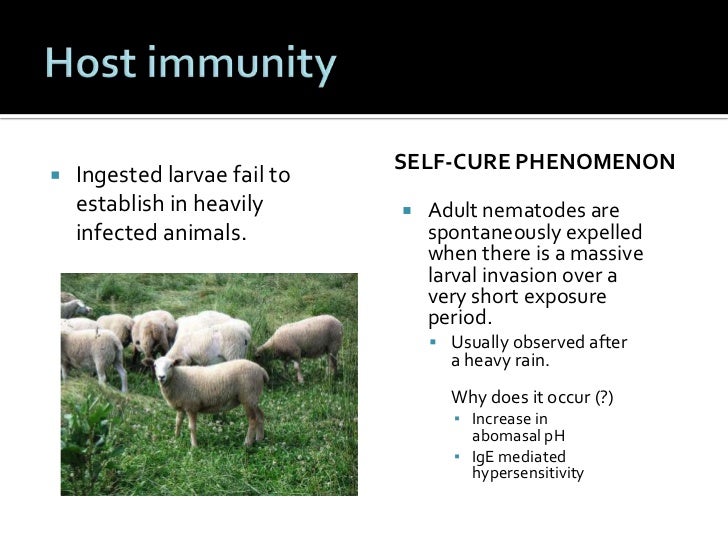
  1. Is an immunological phenomenon in haemonchosis in endemic areas in which as a result of this phenomenon adult worms are expelled out.                                                  
  2. This phenomenon is induced by newly entering infective larvae (L3) in sensitised sheep.                                         
  3. This reaction is initiated when newly entered L3 moult to become L4.                                          
  4. The self cure phenomenon commonly occurs in sheep which have several previous exposure to this parasite.                                                                                        
  5. It doesn't occur in sheep which carry an initial infection (first exposure).                         
  6. It is an immediate type of hypersensitivity reaction and minimum period of 6-7 weeks is required between initial infection and challenge infection for initiating self cure.                                                                                                                                           
  7. In self cure phenomenon, there is a transient rise in blood histamine level, complement fixing antibody and intense mucosal edema.                                               
  8. This phenomenon is not species specific. A challenge infection of H.contortus may expel the adult worm of trichostrongyles.


For more Notes related to parasitology please visit  http://www.univnotes.com/search/label/Parasitolgy

image of a woman and dogA zoonotic disease is a disease that can be spread between animals and humans. Zoonotic diseases can be caused by viruses, bacteria, parasites, and fungi. These diseases are very common. Scientists estimate that more than 6 out of every 10 infectious diseases in humans are spread from animals.
Many people interact with animals in their daily lives. We raise animals for food and keep them in our homes as pets. We might come into close contact with animals at a county fair or petting zoo or encounter wildlife when we clear wooded land for new construction.
Because of these interactions, it’s important to be aware of the different ways people can get zoonotic diseases. These can include:
  • Coming into contact with the saliva, blood, urine, or feces of an infected animal
  • Being bitten by a tick or mosquito (often called a “vector”)
  • Eating or drinking something unsafe (such as unpasteurized milk, undercooked meat, or unwashed fruits and vegetables that are contaminated with feces from an infected animal)
Luckily, there are many ways you can protect yourself and your family from zoonotic diseases. You can:
  • Always wash hands and follow proper hygiene
  • Handle food safely
  • Prevent bites from mosquitoes and ticks
  • Know the simple things you can do to stay safe around your pets
  • Be aware of zoonotic diseases both at home and when you travel

Presentations with additional information for veterinarians and physicians are available in the Resources section. These presentations have been developed to assist in discussions about zoonotic diseases and One Health.

Immunoglobulin M (IgM), the largest pentameric immunoglobulin is used as a marker of recent primary infection for some diseases. IgM constitutes 5-10% of serum immunoglobulins in adults. IgM has half life of 5 days and can be produced in T independent manner.

Structure

There are two forms of IgM
  • Monomeric IgM: Expressed as membrane bound antibody on B cells.
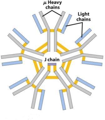
  • Pentameric IgM: Pentameric IgM is secreted by plasma cells. Five monomer subunits are arranged with their Fc regions in the center and ten antigen binding sites on the periphery.
    Pentameric IgM Antibody
    Pentameric IgM Antibody
    The monomers are held together by the interconnection formed in Fc region by disulfide bonds and J (joining chain). J chain, which is added just before secretion of pentamer helps in the polymerization of the monomers.
Properties, Functions and Clinical Significance of IgM
  1. IgM is the earliest immunoglobulin to be synthesized by the fetus beginning by about 20 weeks of age. IgM can not cross the placenta.
    • The presence of IgM in the fetus or newborn indicates intrauterine infection and its detection is useful in the diagnosis of congenital infections such as Toxoplasmosis, syphilis, rubella, cytomegalovirus infection and HIV infection.
  2. IgM is the first immunoglobulin class produced in a primary response to an antigen. IgM antibodies are relatively short lived, disappearing earlier than IgG.
    • Presence of specific IgM against a pathogen in the serum of patients indicates recent/acute infection and helps in disease diagnosis. IgM ELISA test can be performed to detect such antibodies in the serum of patients.
  3. Pentameric IgM has 10 antigen binding sites and 5 Fc portions held together by disulfide bridge bonds and J chains
    • IgM has highest avidity of all immunoglobulins for antigens with multiple epitopes.
    • Less IgM than IgG is required to neutralize viral infectivity. IgM is highly effective against viruses.
    • IgM is a better agglutinin than other antibody isotypes. It takes 100 to 1000 times more molecules of IgG than of IgM to achieve the same level of agglutination.
    • IgM is more efficient than IgG in activating classical pathway of complement. Complement activation requires two Fc regions in close proximity, and the pentameric nature of a single molecule of IgM fulfills this requirement.
  4. Monomeric IgM is the major antibody receptor on the surface of B lymphocytes for antigen recognition.
  5. Large Size: Because of its large size, IgM remains largely confined to bloodstream (80%).  IgM does not diffuse well and therefore is found in very low concentration in the intercellular tissue fluids.
    • IgM is believed to be responsible for protection against blood invasion by microorganisms. IgM deficiency is often associated with septicemias.
  6. High Molecular Weight: Because of its high molecular weight (900,000- 1000,000), it is often called macroglobulin and the ‘millionaire molecule’.
  7. Presence of J chains
    • The presence of J chain allows IgM to bind to receptors on secretory cells, which transport it across epithelial linings to enter the external secretions that bathe mucosal surfaces. Although IgA is the major isotype found in these secretions, IgM plays an important accessory role as a secretory immunoglobulin

pathology এর চিত্র ফলাফলMode of Transmission
Kala-azar is transmitted from person to person by the bite of the female phlebotamine sandfly, p.argentipes which is a highly anthrophilic species. Transmission may also take place by contamination of the bite wound or by contact when the insect is crushed during the act of feeding. Transmission of Kala-azar has also been recorded by blood transfusion, veneral and transplacental but are very rare.
Different factors involved in the transmission of kala-azar
Agent Factor:
  1. Agents: Leishmania donovani, are intracellular parasites that infect and divide within macrophages is the causative agent of kala-azar.
  2. Reservoir of infection: There is a variety of animal reservoirs e.g. dogs, jackals, foxes, rodents and other mammals. Indian kala-azar is considered to be a non-zoonotic infection with man as the sole reservoir.
Host Factor:
  1. Age: Kala-azar can occur in all age groups including infants below the age of one year.
  2. Sex: Males are affected twice as often as females.
  3. Population movement: Movement of Population between endemic and non-endemic areas can result in the spread of infection.
  4. Socio-economic movement: Kala-azar usually affects the poorest of the poor.
  5. Occupation: The disease strongly associated with occupation. People who work in various farming practice, forestry, mining and fishing have a great risk of being bitten by sandflies.
Environmental Factors
  1. Altitudes: Kala-azar is mostly confined to the plains, it does not occur in altitudes over 2000 feet.
  2. Season: High Prevalence during and after rains.
  3. Rural areas: The disease is generally confined to rural areas, where conditions for the breeding of sandflies readily exist compared to urban areas.
  4. Vector: Only the female sandfly transmits the protozoa, infecting itself with the Leishmania parasites contained in the blood it sucks from its human or mammalian host. Sand fly breed in cracks and crevices in the soil and buildings, tree holes, caves etc.


Symptoms

  • Most people infected with Zika virus won’t even know they have the disease because they won’t have symptoms. The most common symptoms of Zika are fever, rash, joint pain, or conjunctivitis (red eyes). Other common symptoms include muscle pain and headache. The incubation period (the time from exposure to symptoms) for Zika virus disease is not known, but is likely to be a few days to a week.
    • See your healthcare provider if you are pregnant and develop a fever, rash, joint pain, or red eyes within 2 weeks after traveling to a place where Zika has been reported. Be sure to tell your health care provider where you traveled.
  • The illness is usually mild with symptoms lasting for several days to a week after being bitten by an infected mosquito.
  • People usually don’t get sick enough to go to the hospital, and they very rarely die of Zika. For this reason, many people might not realize they have been infected.
  • Zika virus usually remains in the blood of an infected person for about a week but it can be found longer in some people.
  • Once a person has been infected, he or she is likely to be protected from future infections.

pathology এর চিত্র ফলাফল Diagnosis

  • The symptoms of Zika are similar to those of dengue and chikungunya, diseases spread through the same mosquitoes that transmit Zika.
  • See your healthcare provider if you develop the symptoms described above and have visited an area where Zika is found.
  • If you have recently traveled, tell your healthcare provider when and where you traveled.
  • Your healthcare provider may order blood tests to look for Zika or other similar viruses like dengue or chikungunya.

Treatment

  • There is no vaccine to prevent or medicine to treat Zika infections.
  • Treat the symptoms:
    • Get plenty of rest.
    • Drink fluids to prevent dehydration.
    • Take medicine such as acetaminophen (Tylenol®) or paracetamol to relieve fever and pain.
    • Do not take aspirin and other non-steroidal anti-inflammatory drugs.
    • If you are taking medicine for another medical condition, talk to your healthcare provider before taking additional medication.
  • If you have Zika, prevent mosquito bites for the first week of your illness.
    • During the first week of infection, Zika virus can be found in the blood and passed from an infected person to a mosquito through mosquito bites.
    • An infected mosquito can then spread the virus to other people.

Powered by Blogger.

Visitors

Print Friendly Version of this pagePrint Get a PDF version of this webpagePDF


 download University Notes apps for android

Popular Posts

Flag Counter