Showing posts with label Anatomy. Show all posts
Why Atlas and Axis are called deviant vertebra?
The vertebra which contains body,archand processes is called a typical vertebra.It has 3 processes.
                                        1.Spinous process
                                        2.Articular process
                                        3.Transverse process
Articular processes are 2 not far away and wide off from each cranial and caudal aspects.
In atlas,the body and spinous process are absent.Theres a fovea dentis which is absent in typical vertebra.Again in axis,the cranial articular orocesses are absent.It has a projection known as odontoid process which is absent in typical vertebra.Thats why atlas and axis are called blinking vertebra.


Description of a typical rib:
Typical Ribs. The typical rib consists of a head, neck and body: The head is wedge shaped, and has two articular facets separated by a wedge of bone. One facet articulates with the numerically corresponding vertebrae, and the other articulates with the vertebrae above.

#A typical rib has a head at the proximal ensue less a costal cartilage at the distal subside
#The shaft is curved and presents two surfaces and two borders
#The lateral surface is convex and has a broad groove at its upper share.
#The medial surface is serene and concave
#Cranial be stuffy to is thick and concave
#Caudal partner occurring is convex
#The head presents 2 articular facets separtes by a groove


Largest foramen of skull:
Image result for Largest foramen of skullforamen magnum is the largest foramen of skull located to the posterior cranial fossa of the occipital bone.
Contents:Vertebral arteries,vertebral nerve.
List the bones of the appendicular skeleton:Appendicular skeleton is made up of the bones of the limbs.
Pectoral limb:Pectoral girdle(shoulder girdle),scapula,clavicle,coracoid,humerus-arm,radius-foramen,ulna-forearm,metacarpal-cannon,phalanges-digits.

Pelvic limb:Pelvic girdle(os coxae),pelvis,ilium,ischium,pubis,femur-thigh,metatarsus cannon,phalanges digits.

Pelvic girdle/bony cavities:
Image result for Pelvic girdle/bony cavitiesIt is formed by two os-coxae(illium+ischium+pubis)or hip bones,sacrum and first few caudal vertebrae.
Ossa-coxarum=os-coxae+os-coxae.
Pelvic inlet: It is formed-
*Dorsally by cranial border of sacrum
*Laterally by shaft of Ilium
*Ventrally cranial border of pubic bone.
Thoracic inlet:Is formed-
*Dorsally by first thoracic vertebra
*Laterally by first pair of ribs
*Ventrally by manubrium of sternum
Thorasic case/bony thorax:is formed-
*Dorsally by series of thorasic vertebra
*Laterally by 13 pairs of ribs

*Ventrally by sternum

Blood supplyLongitudinal section of a digit showing the dorsal

An abundantly rich arterial and capillary vascular network supplies the nail unit. The ulnar and posterior tibial arteries divide into the proper palmar and plantar digital arteries, respectively. The two digital arteries, which occupy the lateral and medial aspects of the digit, form a cruciate anastomosis at the level of the deep dermis in the ventral pulp space. Upon anastomosing on the volar aspect of the terminal phalanx, the two arteries contribute branches to a superficial arcade and the underlying phalangeal periosteum.  The superficial arterial arcade, which also receives a contribution from a vessel coursing dorsally over the DIP joint, supplies the PNF, including the capillary loops of the nail fold and the nail matrix.
The digital arteries continue around the waist of the terminal phalanx and onto the dorsal surface where they divide near the DIP joint to form the dorsal and ventral arches supplying the nail bed and the nail matrix. The dorsal arch courses dorsally over the DIP joint where it terminates in an anastomosis with the proximal arch to supply the matrix and the PNF. The capillary loop system in the PNF is derived from the anastomosis between the two arches.  Therefore, perfusion of the nail matrix comes from two arterial systems: the superficial arcade and the proximal arcade. Anatomically, the superficial arcade sits at the base of the PNF, while the proximal arcade lies deep to the matrix.  The ventral or distal branch supplies the remainder of the nail unit, specifically its most distal aspect, including the fingertips.
An accessory blood supply that is separate from the main blood supply in the pulp space also exists. This accessory system is important in providing adequate perfusion of the nail unit for normal nail growth when the primary vasculature is compromised by disease, which sometimes occurs in scleroderma or infection.
Specialized neurovascular structures called glomus bodies are widely distributed throughout the arterial and deep venous circulation. These oval bodies are encapsulated, consisting of tortuous blood vessels and a neural supply of mostly cholinergic nerves. The glomus bodies form an extensive arteriovenous (AV) anastomotic network, which functions in modulating the thermoregulatory response of the nail unit. Under circumstances where the nail is exposed to cold temperatures, the glomus bodies enhance blood flow to the nail complex by dilating and shunting blood through their complex AV anastomotic systems to prevent ischemia.
The nail apparatus is also richly innervated by a complex neural system allowing tactile sensitivity and sensory discrimination. Anatomically, the laterally positioned digital nerves and their divisions nearly parallel the vascular supply to the nail unit.

The nail plate, the largest segment of the nail unit, is a dense, keratinous horny structure derived from the germinal matrix, or nail root.Destruction of the nail root, despite an intact nail bed, results in permanent loss of the nail plate. Nail growth occurs in a forward distal direction, and growth of the nail plate is directly proportional to the turnover rate of cells in the matrix. Complete growth of the fingernail plate, measured from the matrix to the distal free edge of the plate, is estimated to occur every 4-6 months, corresponding to a rate of 0.1 mm daily.The turnover time for the toenail plate is longer, averaging around 12-18 months.
See the image below.
Longitudinal section of a digit showing the dorsalLongitudinal section of a digit showing the dorsal nail apparatus (top). Component part of the nail apparatus (bottom).
Grossly, the nail plate is composed of 3 distinct layers: a dorsal thin layer, a thick intermediate layer, and a ventral layer. The nail plate originates beneath the PNF and can be seen passing over the lunula.and the richly vascularized nail bed where it firmly attaches. The nail plate is bordered by the PNF and LNFs, and it is distally adjoined by the hyponychium, although its free distal border generally extends beyond the distal phalanx. The healthy nail plate appears as a smooth, convex, nearly rectangular, translucent structure that has a pink color when light is reflected from its surface; the color is due to the underlying vascular-enriched nail bed.
The shape of the nail plate largely conforms to the convexity of the distal margin of the lunula, the visible crescent-shaped distal segment of the matrix. The nail plate exhibits a thickness gradient with the nail thickness, increasing proximally to distally and with age. Thus, the nail plate tends to be thinner at the lunula. Nail thickness is directly related to the length of the matrix.In darkly pigmented persons, a hyperplasia of melanocytes is commonly seen throughout the matrix epithelium, resulting in longitudinal linear bands of light brown to black pigment visible through the nail plate. Although the linear bands of pigment may represent pathological states, they may be a normal finding in people of color, secondary to hyperproduction and accumulation of melanin.
The onychodermal band is the narrow, transverse delineation at the distal end of the nail plate. This band appears yellowish white in whites or reddish brown in darkly pigmented persons. The onychodermal band marks the point of separation of the nail bed from the nail plate,  and it appears to be important in preventing onycholysis after trauma. This band may be prominent in cirrhosis, acrocyanosis, and other chronic diseases. The onychodermal band has been postulated to have a separate vascular supply from the rest of the nail bed

Spinal nerves
Thirty one pairs of spinal nerves exist: 8 cervical, 12 thoracic, 5 lumbar, 5 sacral, and 1 coccygeal.
  • Proximal branches: Each spinal nerve branches into a posterior root and an anterior root. The spinal or posterior root ganglion is occupied by cell bodies from afferent neurons. The convergence of posterior and anterior roots forms the spinal nerve. The cauda equina is formed by the roots arising from segments L2 to Co of the spinal cord.
  • Distal branches: After emerging from the vertebral column, the spinal nerve divides into a posterior ramus, an anterior ramus, and a small meningeal branch that leads to the meninges and vertebral column. The posterior ramus innervates the muscles and joints of the spine and the skin of the back. The anterior ramus innervates the anterior and lateral skin and muscles of the trunk, plus gives rise to nerves leading to the limbs (see image below).
  • Click to see the PDF chart: Nerve and nerve root distribution of major muscles.
  • Nerve plexuses: The anterior rami merge to form nerve plexuses in all areas except the thoracic region (see the image below).
    Unlabelled brachial plexus (for studying). Unlabelled brachial plexus (for studying).
  • Cutaneous innervation and dermatomes: Each spinal nerve except C1 receives sensory input from a specific area of the skin called a dermatome. A dermatome map is a diagram of the cutaneous regions innervated by the branches of each spinal nerve, such a map is an oversimplification, however – each of you is unique as to what you feel.

Image result for Cranial nervesThe cranial nerves emerge from the base of the brain and lead to muscles and sense organs in the head and neck for the most part. The twelve pairs of cranial nerves with their functions are as follows:
  • Olfactory nerve (I): Sensory nerve that carries impulses for smell to the brain.
  • Optic nerve (II): Sensory nerve that carries impulses for vision to the brain.
  • Oculomotor nerve (III): Motor nerve that carries impulses to the extrinsic eye muscles, which help direct the position of the eyeball. This nerve also carries impulses to the muscles that regulate the size of the pupil.
  • Trochlear nerve (IV): Motor nerve that carries impulses to one extrinsic eye muscle (the superior oblique muscle). Once again, this muscle helps regulate the position of the eyeball.
  • Trigeminal nerve (V): A mixed nerve. The sensory fibers of this nerve carry impulses for general sensation (touch, temperature and pain) associated with the face, teeth, lips and eyelids. The motor fibers of this nerve carry impulses to some of the mastication muscles of the face.
  • Abducens nerve (VI): A mixed nerve, but primarily a motor nerve. This nerve carries impulses to the lateral rectus muscle of the eye. This muscle is an extrinsic eye muscle that is involved in positioning the eyeball.
  • Facial nerve (VII): A mixed nerve. The sensory fibers of this nerve carry taste sensations from the tongue. The motor fibers of this nerve carry impulses to many of the muscles of the face and they carry impulses to the lacrimal, submandibular, and sublingual glands.
  • Vestibulocochlear nerve (VIII): A sensory nerve that carries impulses for hearing and equilibrium from the ear to the brain.
  • Glossopharyngeal nerve (IX): A mixed nerve. The sensory fibers of this nerve carry basic sensory information and taste sensations from the pharynx and tongue to the brain. The motor fibers of this nerve carry impulses associated with swallowing to the pharynx.
  • Vagus nerve (X): A mixed nerve. The sensory fibers of this nerve carry impulses from the pharynx, larynx, and most internal organs to the brain. The motor fibers of this nerve carry impulses to internal organs of the chest and abdomen and to the skeletal muscles of the larynx and pharynx.
  • Accessory nerve (XI): A mixed nerve, but primarily motor. Carries impulses to muscles of the neck and back.
  • Hypoglossal nerve (XII): Primarily a motor nerve. This nerve carries impulses to the muscles that move and position the tongue.


The sensory (afferent) division carries sensory signals by way of afferent nerve fibers from receptors in the central nervous system (CNS). It can be further subdivided into somatic and visceral divisions. The somatic sensory division carries signals from receptors in the skin, muscles, bones and joints. The visceral sensory division carries signals mainly from the viscera of the thoracic and abdominal cavities.
The motor (efferent) division carries motor signals by way of efferent nerve fibers from the CNS to effectors (mainly glands and muscles). It can be further subdivided into somatic and visceral divisions. The somatic motor division carries signals to the skeletal muscles. The visceral motor division, also known as the autonomic nervous system, carries signals to glands, cardiac muscle, and smooth muscle. It can be further divided into the sympathetic and parasympathetic divisions.
The sympathetic division tends to arouse the body to action. The parasympathetic divisions tend to have a calming effect.
Nerve fibers of the PNS are classified according to their involvement in motor or sensory, somatic or visceral pathways. Mixed nerves contain both motor and sensory fibers. Sensory nerves contain mostly sensory fibers; they are less common and include the optic and olfactory nerves. Motor nerves contain motor fibers.
Peripheral nerve supply to the muscles in the lowePeripheral nerve supply to the muscles in the lower extremity.
Peripheral nerve supply to the muscles in the uppePeripheral nerve supply to the muscles in the upper extremity.

The peripheral nervous system refers to parts of the nervous system outside the brain and spinal cord. It includes the cranial nerves, spinal nerves and their roots and branches, peripheral nerves, and neuro-muscular junctions. The anterior horn cells, although technically part of the central nervous system (CNS), are sometimes discussed with the peripheral nervous system because they are part of the motor unit.
Bundles of nerve fibers or axons conduct information to and from the central nervous system. The autonomic nervous system is the part of the nervous system concerned with the innervation of involuntary structures, such as the heart, smooth muscle, and glands within the body. It is distributed throughout the central and peripheral nervous systems.
An image depicting the peripheral nervous system can be seen below.
Major divisions of the peripheral nervous system. Major divisions of the peripheral nervous system.
Nerve cells are called neurons. A neuron consists of a cell body (with a nucleus and cytoplasm), dendrites that carry electrical impulses to the cell, and a long axon that carries the impulses away from the cell. The axon of one neuron and the dendrites of the next neuron do not actually touch. The gap between neurons is called the synapse.
Neuronal function is complex and involves numerous processes in nerve transmission. Generation of a nerve impulse (action potential) of a sensory neurons occurs as a result of a stimulus such as light, a particular chemical, or stretching of a cell membrane by sound. Conduction of an impulse along a neuron occurs from the dendrites to the cell body to the axon. Transmission of a signal to another neuron across a synapse occurs via chemical transmitter. This substance causes the next neuron to be electrically stimulated and keeps the signal going along a nerve.
Ganglia may be divided into sensory ganglia of spinal nerves (spinal or posterior root ganglia) and cranial nerves and autonomic ganglia. Sensory ganglia of spinal nerves are fusiform swellings situated on the posterior root of each spinal nerve just proximal to the root’s junction with a corresponding anterior root. They are referred to as spinal or posterior root ganglia. Similar ganglia that are also found along the course of cranial nerves V, VII, VIII, IX, and X are called sensory ganglia of these nerve. Autonomic ganglia, which are often irregular in shape, are situated along the course of efferent nerve fibers of the autonomic nervous system. They are found in the para vertebral sympathetic chains, around the roots of the great visceral arteries in the abdomen, and close to, or embedded within, the walls of various viscera.

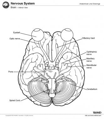
Brain, inferior view.
The neocortex is the most phylogenetically developed structure of the human brain as compared with the brains of other species. The complex pattern of folding allows an increased cortical surface to occupy a smaller cranial volume. The pattern of folding that forms the sulcal and gyral patterns remains highly preserved across individuals. This enables a nomenclature for the cortical anatomy.

The left and right cerebral hemispheres are separated by the longitudinal cerebral fissure. The principal connection between the 2 hemispheres is the corpus callosum. Each cortical hemisphere can be divided into 4 lobes: frontal, temporal, parietal, and occipital. The frontal lobe can be distinguished from the temporal lobe by the lateral sulcus (Sylvian fissure). The frontal lobe can be distinguished from the parietal lobe by the central sulcus (Rolandic fissure). The parieto-occipital sulcus, which is visible on the medial aspect of the hemisphere, divides the parietal and occipital lobes. Within the lateral sulcus is another cortical surface referred to as the insula.
The frontal lobe can then be further divided into the superior, middle, and inferior frontal gyri, which are divided by the superior and inferior frontal sulci, respectively. The inferior frontal gyrus forms the frontal operculum, which overlies the lateral sulcus. The frontal operculum can be divided into 3 triangular gyri: the pars orbitalis, the pars triangularis, and the pars opercularis, in order from anterior to posterior. The precentral gyrus is the gyrus immediately anterior to the central sulcus.
Similarly, the temporal lobe is divided into the superior, middle, and inferior temporal gyri, which are separated by the superior and inferior temporal sulci. On the inferior surface of the temporal lobe just lateral to the midbrain the parahippocampal gyrus can be identified, with the collateral sulcus lying lateral. Between the parahippocampal gyrus and the inferior temporal gyrus lies the occipitotemporal gyrus, also known as the fusiform gyrus.
Within the parietal lobe, the superior temporal sulcus is capped by the angular gyrus. Just above this, the lateral sulcus is capped by the supramarginal gyrus. Just below the angular gyrus, the lateral occipital gyrus caps the inferior temporal sulcus. 


Brain, coronal view. Brain, coronal view.
Brain, inferior view. Brain, inferior view.
Brain, lateral view. Brain, lateral view.
Brain, midsagittal view. Brain, midsagittal view

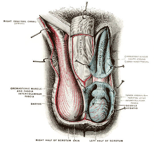
File:Gray1143.png

A.  Functions


1.  Produce male germ cells (spermatozoa)


2.  Produce male hormone (testosterone)


3. Produce inhibin and estrogen, and other proteins


B.  Structure


1.  Tunica vaginalis


a.  Thin membrane


b.  Provides support


c.  From peritoneum


2.  Tunica albuginea


a.  Connective tissue


b.  Provides structure


3.  Mediastinum


a.  Connective tissue


  1. Provides internal support
    4.  Seminiferous tubules
    part of the testicular parenchyma
    a.  Site of sperm production
    b. Consists of 2 cell types 
    - Germ cells (eventual sperm cells)
    - Sertoli or nurse cells which produces variety of substances including androgen binding hormone, sulfated glycoprotein, transferin, and inhibin
    - Surround developing germ cells
    - Providing structural and metabolic support to the developing spermatogenic cells
    3.  Blood-testis Barriers
    a. Cells surrounding the seminiferous tubules
    b. Prevent autoimmune reaction from destroying the developing germ cells
    5.  Interstitial or Leydig cells
    a.  Located between seminiferous tubules
    b.  Produce androgens (testosterone)
    6.  Rete testis
    a.  Collect sperm from seminiferous tubules
    7.  Vasa efferentia (efferent ductules)
    a.  Collect sperm from rete testis
    b.  Carry sperm out of testis proper

Image result for cervical vertebra
The cervical vertebrae of the spine consist of seven bony rings that reside in the neck between the base of the skull and the thoracic vertebrae in the trunk. Among the vertebrae of the spinal column, the cervical vertebrae are the thinnest and most delicate bones. Yet, in spite of their size, the cervical vertebrae have the huge jobs of supporting the head, protecting the spinal cord, and providing mobility to the head and neck.
The cervical vertebrae are stacked along the length of the neck to form a continuous column between the skull and the chest Each cervical vertebra is named by its position in order from superior (C1 or first cervical vertebra) to inferior (C7 or seventh cervical vertebra). The C1 vertebra, which holds up the skull, is named the atlas after the mythological titan Atlas who similarly held the Earth on his shoulders. Similar to the C1 vertebra, the C2 vertebra is named the axis as it provides the axis upon which the skull and atlas rotate when the head is moved side to side.
Each cervical vertebra consists of a thin ring of bone, or vertebral arch, surrounding the vertebral and transverse foramina. The vertebral foramen is a large opening in the center of the vertebra that provides space for the spinal cord and its meninges as they pass through the neck. Flanking the vertebral foramen on each side are the much smallertransverse foramina. The transverse foramina surround the vertebral arteries and veins, which, along with the carotid arteries and jugular veins, have the vital job of carrying blood to and from the brain.
Extending from the vertebral arch are several bony processes that are involved in muscle attachment and movement of the neck. The spinous process extends from the posterior end of the arch and serves as a connection point for the muscles that extend the neck, such as the trapezius and spinalis muscles. On the left and right lateral sides of each vertebra is a transverse process that forms the insertion point for the muscles of the erector spinae group that extend and flex the neck.
A thickened region of bone known as the body lies anterior to the vertebral foramen and forms the main bone mass in all vertebrae except for the atlas. The bodies strengthen the vertebrae and support most of the weight of the tissues of the head and neck.Intervertebral disks made of rubbery fibrocartilage lie between the vertebral bodies to provide slight flexibility to the neck. Lateral to the vertebral bodies are flattened facets that form joints with the neighboring vertebrae and skull, allowing movement among the vertebrae. The axis has a very distinct shape due to the presence of the odontoid process, a tooth-like prominence that extends from its body superiorly toward the axis. The odontoid process serves as the axis upon which the atlas rotates at the atlantoaxial joint.
Despite being some of the smallest and lightest bones in the axial skeleton, the cervical vertebrae perform many important functions that are critical to the survival of the body. Vital nerves and blood vessels passing through the neck are protected from mechanical damage by the bony arches of the cervical vertebrae. The cervical vertebrae also provide support to the head and neck, including supporting the muscles that move this region of the body. The muscles that attach to the vertebral processes provide posture to the head and neck throughout the day and have the greatest endurance of all of the body’s muscles. Finally, the many joints formed between the skull and cervical vertebrae provide incredible flexibility that allows the head and neck to rotate, flex, and extend.

Powered by Blogger.

Visitors

Print Friendly Version of this pagePrint Get a PDF version of this webpagePDF


 download University Notes apps for android

Popular Posts

Flag Counter