
Diagnosis of Visceral Leishmaniasis may require taking a blood
sample and/or taking a biopsy from the bone marrow or splenic puncture
to show the parasite.
- Diagnosis of Cutaneous Leishmaniasis will require a small biopsy or scrapping of ulcer.
- Diagnosis of Mucocutaneous Leishmaniasis requires a biopsy of the affected tissues.
Biopsy samples are examined by microscopy, culture and other
methods to look for the parasite and identify the specific kind of
leishmania causing the ulcer.
Sample for Visceral Leishmaniasis

-
Blood: For making Blood film/Blood Culture and Serological Tests.
-
Bone Marrow: Biopsy material obtained by sternal or iliac crest puncture.
-
Splenic Pulp: Biopsy material obtained by splenic culture.As the clinical presentation of Visceral Leishmanisis (Kala azar) lacks specificity, confirmatory tests are required to decide which patients should be treated. Such tests should be highly sensitive (>95%) as VL is a fatal condition, but also highly specific because the current drugs used to treat VL are toxic. Ideally, a test should be able to make the distinction between acute disease and asymptomatic infection, because none of the drugs currently available is safe enough to treat asymptomatic infections. Moreover, such tests should be simple and affordable.
Non Leishmanial Test:
a. Detection of reduction in the number of red and white blood
cells and platelets (pancytopenia) in VL suspected clinical patients.
This test is highly specific but sensitivity is very low.
b. Aldedhyde Test (also called Formal Gel Test: FGT): Polyclonal
hypergammaglobulinemia (the production of high titres of non-specific
antibody) is detected in this test. Sensitivity of this test is poor, as
low as 34%.
In this test 1-2 drops of serum from a case of kala-azar is taken
and a drop or two of formalin is added. A positive test is indicated by
jellification to milk white opacity like the white of a hard boiled egg
within 3 minute to 24 hours.
Leishmanin Test:
A killed culture (0.1-0.2 ml) of a suspension containing 6 to 10
promastigotes per ml is injected intradermally. A positive reaction (an
area of induration after 72 hours) is produced in cured kala azar cases 6
to 8 weeks after recovery and represent a delayed hypersensitivity
reaction accompanied by cell-mediated immunity. The test is positive in
African Kala azar but not in indian and mediterranean kala-azar and post
kala azar dermal leishmaniasis.

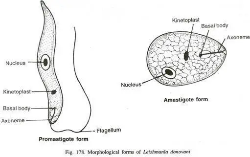
Morphological form of Leishmania donovani
Parasite detection
For the detection of parasite visualization of the amastigote form
of the parasite by microscopic examination of aspirates from lymph
nodes, bone marrow, or spleen is done. It is the classic confirmatory
test for Visceral Leishmaniasis.
Although the specificity is high, the sensitivity of microscopy varies, being higher for spleen (93% to 99%) than for bone marrow (53-86%) or lymph nodes aspirates (53-65%) .
Although the specificity is high, the sensitivity of microscopy varies, being higher for spleen (93% to 99%) than for bone marrow (53-86%) or lymph nodes aspirates (53-65%) .
The detection of parasites in the blood or organs by culture or by
using molecular techniques such as PCR is more sensitive than
microscopic examination.


0 Comments:
Post a Comment