
 Main Diseases caused by Staphylococcus aureus
- Abscesses
- Endocarditis
- Gastroenteritis (food poisoning)
- Toxic Shock Syndrome
- Hospital Acquired Pneumonia
- Sepsis
- Surgical wound infections
- Staphylococcal scalded skin syndrome (SSSS)
Important Properties of Staphylococcus aureus
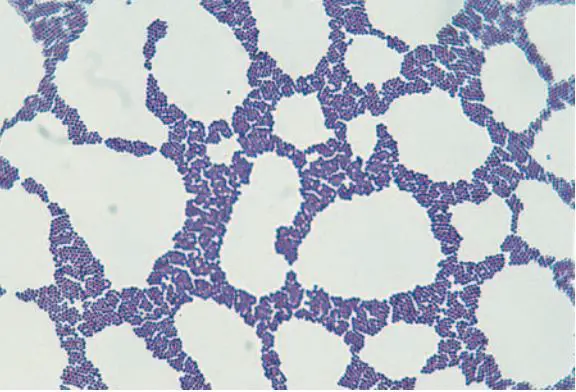
- Gram positive cocci that occur singly and in pairs, tetrads, short chains, and irregular grapelike clusters
Staphylococcus in Gram Stain
- Catalase Test: positive
- Coagulase Test: positive
- Other properties: Non motile, non sporing, often unencapsulated or have a limited capsule, facultative anaerobes.
- Surface proteins: Protein A (prevents activation of Complement), Clumping factor, Teichoic acid (adherence and induction of septic shock)
- Super antigens: Enterotoxin A-D, Exfoliatin A&B, Toxic Shock Syndrome Toxin (TSST)-super antigen.
- Cytotoxins: α-hemolysin, β-hemolysin,γ-hemolysin, δ-hemolysin, Panton Valentine Leukocidin (PVL), Exfliatin (epidermolytic-cleaves desmglein in desmosomes)
- Enzymes: Nuclease, Lipase, Hyaluronidase, Coagulase (causes plasma to clot), Staphylokinase, Glyerol ester hydrolase, Catalase (degradation of H2O2 limits the ability of neutrophils to kill S. aurues), etc.
- Gram staining: Gram positive cocci in clusters is the characteristics, cocci may appear singly in pairs or in short chains.
- Culture:
- Blood Agar– Colony morphology: growth occurs abundantly within 18 to 24 hours, yellow or golden yellow colonies with or without Beta hemolysis are seen.
- Mannitol Salt Agar (MSA) is commonly used to screen S. aureus: Gives yellow colored colonies.
- Biochemical tests:
- Catalase test: Positive
- Coagulase test: Positive- Distinguish S. aureus from Coagulase Negative Staphylococcus aureus (CONS). CONS are further differentiated on the basis of Novobiocin sensitivity test ( S. epidermidis is sensitive, whereas S. saprophyticus is resistant).


0 Comments:
Post a Comment