
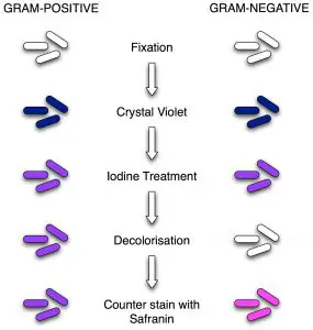
 We all know that Gram stain is the most important staining
technique for identifying bacteria using light microscopy but Gram
staining techniques also have some limitations. We know how to perform
gram stain but we may not know the procedure we applied is adequate or
not.
We all know that Gram stain is the most important staining
technique for identifying bacteria using light microscopy but Gram
staining techniques also have some limitations. We know how to perform
gram stain but we may not know the procedure we applied is adequate or
not.- Limitation of Gram Stain: Mycobacteria stain weakly with gram stain and bacteria such as Mycoplasma, Rickettsiae, Chlamydiae do not take up the dyes used in Gram stain or are too small to be seen with light microscopy.
- Sensitivity of Gram Staining Technique: To be visible on a slide, organisms that stain by the Gram method must be present in concentrations of about 10^4 to 10^5 organisms per milliliter of uncentrifuged fluid.
- Adequacy of Gram Staining method: After performing gram stain, Microbiologist/Technician should first determine whether the Gram stain is adequate. In an appropriately stained specimen, the nuclei of neutrophils are red. If the nuclei are blue, the decolorization is insufficient.
- Gram positive bacteria may lose their ability to retain crystal violet and stain Gram negatively for the following reasons:
- Cell wall damage of bacteria due to antibiotic therapy or excessive heat fixation of the smear.
- Over- decolorization of the smear
- Use of an Iodine solution which is too old, i.e. yellow instead of brown in color (always store in a brown glass or other light opaque container).
- Smear has been prepared from an old culture.
- When smear is too thick, Gram negative bacteria may not be fully decolorized during decolorization steps and appear as Gram positive bacteria.


0 Comments:
Post a Comment