M. leprae in stained smear Sample: Skin lesions or nasal scrapings or specimen from ear lobules.
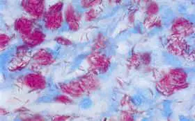
a.  Microscopy (Slit-skin smear stained with modified Ziehl Neelsen stain: 4 to 5% Sulphuric acid as decolourising agent)
M. leprae in stained smear
  • For Lepromatous leprosy: lipid laden macrophages called ‘foam cells” containing many acid fast bacilli are seen in the skin.
  • For Tuberculoid leprosy: Very few acid fast bacilli are seen and appearance of typical granulomas is sufficient for diagnosis.
M. leprae are slightly curved filament 3-10 m in length containing irregular arrangements of dense material sometimes in the shape of rods. M. leprae which stain with carbol-fuchsin as solid acid-fast rods are believed to be viable (not viable in smear now but in the host body during that time) and that bacilli which stain irregularly are probably dead and degenerating.
Based on the number of of M. lepare and their morphology in the stained slides Bacteriological index (BI) and  morphological index (MI) can be calculated. These indices are useful in assessing the amount of infection, viability of the organisms and also the progress of the patient under treatment.  Bacteriological index (BI) is an expression of the extent of bacterial loads Where as morphological index (MI) is calculated by counting the numbers of solid-staining acid-fast rods. According to WHO, a more accurate and reliable index of the bacillary content of a lesion is given by the logarithmic index of biopsies (LIB) is a more accurate and reliable index of the bacillary content of a lesion. These indices help to assess the state of patients at the beginning of treatment and to assess progress.

b. Culture
M. leprae has not yet been successfully cultured in vitro but it can be grown in the laboratory by injection into the foot pads of mice or nine-banded armadillo. It is a slow growing pathogen with the doubling time of 14 days.
c. Molecular diagnosis: Polymerase Chain Reaction (PCR) can be used as a means of diagnosis of leprosy and also as a tool for  drug assessment.

Related Posts:

  • Catalase, Oxidase & Coagulase test Catalase Test This test is used to identify organisms that produce the enzyme, catalase. This enzyme detoxifies hydrogen peroxide by breaking it down into water and oxygen gas. … Read More
  • Streak-stab technique & MacConkey agar Streak-stab technique Often when inoculating a BAP to observe hemoloysis patterns, investigators will also stab several times through the agar using an inoculating loop. This … Read More
  • SIM media Sulfur Indole Motility Media (SIM) This is a differential medium. It tests the ability of an organism to do several things: reduce sulfur, produce indole and swim through … Read More
  • Nitrate Broth This is a differential medium. It is used to determine if an organism is capable of reducing nitrate (NO3-) to nitrite (NO2-) or other nitrogenous compounds via the action … Read More
  • Kliger’s Iron Agar (KIA) Kliger’s Iron Agar (KIA) This is a differential medium. It tests for organisms’ abilities to ferment glucose and lactose to acid and acid plus gas end products. It also allows for ident… Read More

0 Comments:

Powered by Blogger.

Visitors

220619
Print Friendly Version of this pagePrint Get a PDF version of this webpagePDF


 download University Notes apps for android

Popular Posts

Flag Counter