
The nail plate, the largest segment of the nail unit, is a dense, keratinous horny structure derived from the germinal matrix, or nail root.Destruction of the nail root, despite an intact nail bed, results in permanent loss of the nail plate. Nail growth occurs in a forward distal direction, and growth of the nail plate is directly proportional to the turnover rate of cells in the matrix. Complete growth of the fingernail plate, measured from the matrix to the distal free edge of the plate, is estimated to occur every 4-6 months, corresponding to a rate of 0.1 mm daily.The turnover time for the toenail plate is longer, averaging around 12-18 months.
See the image below.
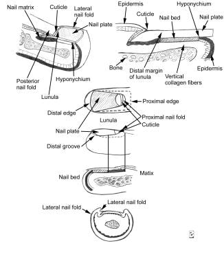 Longitudinal section of a digit showing the dorsal nail apparatus (top). Component part of the nail apparatus (bottom).
Grossly, the nail plate is composed of 3 distinct layers: a dorsal thin layer, a thick intermediate layer, and a ventral layer. The nail plate originates beneath the PNF and can be seen passing over the lunula.and the richly vascularized nail bed where it firmly attaches. The nail plate is bordered by the PNF and LNFs, and it is distally adjoined by the hyponychium, although its free distal border generally extends beyond the distal phalanx. The healthy nail plate appears as a smooth, convex, nearly rectangular, translucent structure that has a pink color when light is reflected from its surface; the color is due to the underlying vascular-enriched nail bed.
The shape of the nail plate largely conforms to the convexity of the distal margin of the lunula, the visible crescent-shaped distal segment of the matrix. The nail plate exhibits a thickness gradient with the nail thickness, increasing proximally to distally and with age. Thus, the nail plate tends to be thinner at the lunula. Nail thickness is directly related to the length of the matrix.In darkly pigmented persons, a hyperplasia of melanocytes is commonly seen throughout the matrix epithelium, resulting in longitudinal linear bands of light brown to black pigment visible through the nail plate. Although the linear bands of pigment may represent pathological states, they may be a normal finding in people of color, secondary to hyperproduction and accumulation of melanin.
The onychodermal band is the narrow, transverse delineation at the distal end of the nail plate. This band appears yellowish white in whites or reddish brown in darkly pigmented persons. The onychodermal band marks the point of separation of the nail bed from the nail plate, and it appears to be important in preventing onycholysis after trauma. This band may be prominent in cirrhosis, acrocyanosis, and other chronic diseases. The onychodermal band has been postulated to have a separate vascular supply from the rest of the nail bed

0 Comments:
Post a Comment